製品: ACP5 Antibody
カタログ: DF6989
タンパク質の説明: Rabbit polyclonal antibody to ACP5
アプリケーション: WB IHC
反応性: Human, Mouse, Rat
予測: Pig, Bovine, Horse, Sheep, Rabbit, Dog
分子量: 37kDa; 37kD(Calculated).
ユニプロット: P13686
RRID: AB_2838945

類似製品を見る>>

   サイズ 価格 在庫状況
 100ul $280 在庫あり
 200ul $350 在庫あり

リードタイム: 当日配達

For pricing and ordering contact:
お問い合わせ先

製品説明

ソース:
Rabbit
アプリケーション:
WB 1:500-1:2000, IHC 1:50-1:200
*The optimal dilutions should be determined by the end user.
*Tips:

WB: For western blot detection of denatured protein samples. IHC: For immunohistochemical detection of paraffin sections (IHC-p) or frozen sections (IHC-f) of tissue samples. IF/ICC: For immunofluorescence detection of cell samples. ELISA(peptide): For ELISA detection of antigenic peptide.

反応性:
Human,Mouse,Rat
予測:
Pig(100%), Bovine(92%), Horse(92%), Sheep(92%), Rabbit(100%), Dog(92%)
クローナリティ:
Polyclonal
特異性:
ACP5 Antibody detects endogenous levels of total ACP5.
RRID:
AB_2838945
引用形式: Affinity Biosciences Cat# DF6989, RRID:AB_2838945.
コンジュゲート:
Unconjugated.
精製:
The antiserum was purified by peptide affinity chromatography using SulfoLink™ Coupling Resin (Thermo Fisher Scientific).
保存:
Rabbit IgG in phosphate buffered saline , pH 7.4, 150mM NaCl, 0.02% sodium azide and 50% glycerol. Store at -20 °C. Stable for 12 months from date of receipt.
別名:

折りたたみ/展開

Type 5 acid phosphatase; Acid phosphatase 5 tartrate resistant; Acid phosphatase 5, tartrate resistant; ACP5; EC 3.1.3.2; MGC117378; PPA5_HUMAN; serum band 5 tartrate-resistant acid phosphatase; SPENCDI; T5ap; Tartrate resistant acid ATPase; Tartrate resistant acid phosphatase type 5; Tartrate resistant acid phosphatase type 5 precursor; Tartrate-resistant acid ATPase; Tartrate-resistant acid phosphatase type 5; TR AP; TR-AP; TRACP 5; TRAP; TrATPase; Type 5 acid phosphatase;

免疫原

免疫原:
Uniprot:
遺伝子(ID):
タンパク質の説明:
This gene encodes an iron containing glycoprotein which catalyzes the conversion of orthophosphoric monoester to alcohol and orthophosphate. It is the most basic of the acid phosphatases and is the only form not inhibited by L(+)-tartrate.
タンパク質配列:
MDMWTALLILQALLLPSLADGATPALRFVAVGDWGGVPNAPFHTAREMANAKEIARTVQILGADFILSLGDNFYFTGVQDINDKRFQETFEDVFSDRSLRKVPWYVLAGNHDHLGNVSAQIAYSKISKRWNFPSPFYRLHFKIPQTNVSVAIFMLDTVTLCGNSDDFLSQQPERPRDVKLARTQLSWLKKQLAAAREDYVLVAGHYPVWSIAEHGPTHCLVKQLRPLLATYGVTAYLCGHDHNLQYLQDENGVGYVLSGAGNFMDPSKRHQRKVPNGYLRFHYGTEDSLGGFAYVEISSKEMTVTYIEASGKSLFKTRLPRRARP

種類予測

種類予測:

Score>80(red) has high confidence and is suggested to be used for WB detection. *The prediction model is mainly based on the alignment of immunogen sequences, the results are for reference only, not as the basis of quality assurance.

Species
Results
Score
Rabbit
100
Pig
100
Horse
92
Bovine
92
Sheep
92
Dog
92
Chicken
75
Xenopus
58
Zebrafish
0
Model Confidence:
High(score>80) Medium(80>score>50) Low(score<50) No confidence

PTMs - P13686 基板として

Site PTM Type Enzyme
N116 N-Glycosylation
K222 Ubiquitination
Y278 Phosphorylation
Y283 Phosphorylation

研究背景

機能:

Involved in osteopontin/bone sialoprotein dephosphorylation. Its expression seems to increase in certain pathological states such as Gaucher and Hodgkin diseases, the hairy cell, the B-cell, and the T-cell leukemias.

細胞の位置付け:

Lysosome.

Extracellular region or secreted Cytosol Plasma membrane Cytoskeleton Lysosome Endosome Peroxisome ER Golgi apparatus Nucleus Mitochondrion Manual annotation Automatic computational assertionSubcellular location
サブユニット構造:

Exists either as monomer or, after proteolytic processing, as a dimer of two chains linked by disulfide bond(s).

タンパク質ファミリー:

Belongs to the metallophosphoesterase superfamily. Purple acid phosphatase family.

研究領域

· Cellular Processes > Transport and catabolism > Lysosome.   (View pathway)

· Human Diseases > Immune diseases > Rheumatoid arthritis.

· Metabolism > Metabolism of cofactors and vitamins > Riboflavin metabolism.

· Metabolism > Global and overview maps > Metabolic pathways.

· Organismal Systems > Development > Osteoclast differentiation.   (View pathway)

参考文献

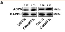
1). RANK promotes colorectal cancer migration and invasion by activating the Ca2+-calcineurin/NFATC1-ACP5 axis. Cell Death & Disease, 2021 (PubMed: 33795653) [IF=8.1]

Application: WB    Species: Human    Sample: SW480 and Caco2 cells

Fig. 3 RANK regulated CRC migration and invasion by activating ACP5 expression. a, b Western blot analysis of upregulation of ACP5 protein in RANK-overexpressing SW480 and Caco2 cells, and downregulation of ACP5 protein in RANK knockdown HT29 cells. c, f Western blotting showed that successful decreased ACP5 protein in RANK overexpression CRC cells transfected with siRNA-ACP5 and increased ACP5 protein by transfection with pFV-ACP5 plasmid in RANK knockdown CRC cells. d, e Knockdown of ACP5 inhibited RANK-induced migration and invasion in SW480RK and Caco2RK cells. Scales bars = 100 μm. g, h ACP5 overexpression increased migration and invasion of RANK-silencing HT29 cells. Scales bars = 100 μm. i Representative immunofluorescence of ACP5 distribution in high and low RANK expression CRC tissues. Nuclei (blue) were counterstained with DAPI. The white arrowheads indicate ACP5-positive cells are distributed within the tumor and stroma with high RANK expression. + (score 1–4), +++ (score 9–12). Scales bars = 10 μm. Data are mean ± SD (n = 3).

Application: IF/ICC    Species: Human    Sample: SW480 and Caco2 cells

Fig. 3 RANK regulated CRC migration and invasion by activating ACP5 expression. a, b Western blot analysis of upregulation of ACP5 protein in RANK-overexpressing SW480 and Caco2 cells, and downregulation of ACP5 protein in RANK knockdown HT29 cells. c, f Western blotting showed that successful decreased ACP5 protein in RANK overexpression CRC cells transfected with siRNA-ACP5 and increased ACP5 protein by transfection with pFV-ACP5 plasmid in RANK knockdown CRC cells. d, e Knockdown of ACP5 inhibited RANK-induced migration and invasion in SW480RK and Caco2RK cells. Scales bars = 100 μm. g, h ACP5 overexpression increased migration and invasion of RANK-silencing HT29 cells. Scales bars = 100 μm. i Representative immunofluorescence of ACP5 distribution in high and low RANK expression CRC tissues. Nuclei (blue) were counterstained with DAPI. The white arrowheads indicate ACP5-positive cells are distributed within the tumor and stroma with high RANK expression. + (score 1–4), +++ (score 9–12). Scales bars = 10 μm. Data are mean ± SD (n = 3).

2). Schistosoma japonicum cystatin suppresses osteoclastogenesis via manipulating the NF‑κB signaling pathway. Molecular Medicine Reports, 2021 (PubMed: 33576450) [IF=3.4]

Application: WB    Species:    Sample: RAW264.7 cells

Figure 4. |rSj‑Cys inhibits the expression of osteoclastogenesis‑related genes and proteins. RAW264.7 cells were co‑stimulated with M‑CSF (25 ng/ml) and RANKL (30 ng/ml) and then treated with or without rSj‑Cys (0.3 µM) for different time periods (24, 48 or 72 h). The expression levels of genes and proteins associated with osteoclast phenotype markers (CTSK, TRAP, ITGB3) and cell surface receptors of osteoclast precursors (RANK, OSCAR, TREM‑2)were determined by (A) reverse transcription‑quantitative PCR and (B) western blot analysis.

3). Xuetongsu attenuates bone destruction in collagen-induced arthritis mice by inhibiting osteoclast differentiation and promoting osteoclast apoptosis. The international journal of biochemistry & cell biology, 2024 (PubMed: 38340949) [IF=3.4]

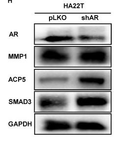
4). Androgen receptor (AR) decreases HCC cells migration and invasion via miR-325/ACP5 signaling. Journal of Cancer, 2023 (PubMed: 33753989) [IF=3.3]

Application: WB    Species: Human    Sample: HCC cells

Figure 1 AR can decrease the progression of HCC cells while ACP5 can increase. A-B. Western blot was used to check AR protein expression after shAR/oeAR in HCC cells. C-D. Wound-healing and transwell invasion assay was used to check the migration and invasion capacity in the HA22T shAR cells. E-F. Wound-healing and transwell invasion assay was used to check the migration and invasion capacity in the SK-HEP-1 oeAR cells. G-H. Western blot assay was used to check related protein expressions after over expression AR/knocking down AR in HCC cells. I-J. Western blot assay was used to check ACP5 expression after shACP5/oeACP5 in HCC cells. K-L. Wound-healing assay was used to check the migration capacity after shACP5/oeACP5 in HCC cells. M-N. Transwell invasion assay was used to check the invasion capacity after shACP5/oeACP5 in HCC cells. All quantifications are mean ± SD, *p < 0.05, **p < 0.01, ***p < 0.001, ns: no significant difference.

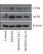
5). Tanshinone I Mitigates Steroid-Induced Osteonecrosis of the Femoral Head and Activates the Nrf2 Signaling Pathway in Rats. Evidence-Based Complementary and Alternative Medicine, 2021 (PubMed: 35111227)

Application: WB    Species: Rat    Sample:

Figure 3 TsI restrains osteoclastogenesis in the femoral heads of the MPS-induced SIONFH rat model. (a) TRAP activity in the serum. (b) Expression levels of CTSK and ACP5 in the femoral heads. ∗∗p < 0.01; ns, no significant. TRAP, tartrate-resistant acid phosphatase; CTSK, cathepsin K; ACP5, acid phosphatase 5.

Restrictive clause


Affinity Biosciences tests all products strictly. Citations are provided as a resource for additional applications that have not been validated by Affinity Biosciences. Please choose the appropriate format for each application and consult Materials and Methods sections for additional details about the use of any product in these publications.

For Research Use Only.
Not for use in diagnostic or therapeutic procedures. Not for resale. Not for distribution without written consent. Affinity Biosciences will not be held responsible for patent infringement or other violations that may occur with the use of our products. Affinity Biosciences, Affinity Biosciences Logo and all other trademarks are the property of Affinity Biosciences LTD.