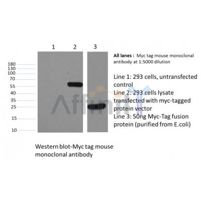
C-Myc-Tag Antibody - #T0001
| 製品: | C-Myc-Tag Antibody |
| カタログ: | T0001 |
| タンパク質の説明: | Mouse monoclonal antibody to C-Myc-Tag |
| アプリケーション: | WB IP ELISA |
| Cited expt.: | WB |
| 反応性: | All |
| ユニプロット: | |
| RRID: | AB_2839411 |
製品説明
ソース:
Mouse
アプリケーション:
WB 1:3000-1:10000, IF/ICC: 1:200, IP 1:200
*The optimal dilutions should be determined by the end user. For optimal experimental results, antibody reuse is not recommended.
*Tips:
*The optimal dilutions should be determined by the end user. For optimal experimental results, antibody reuse is not recommended.
*Tips:
WB: For western blot detection of denatured protein samples. IHC: For immunohistochemical detection of paraffin sections (IHC-p) or frozen sections (IHC-f) of tissue samples. IF/ICC: For immunofluorescence detection of cell samples. ELISA(peptide): For ELISA detection of antigenic peptide.
反応性:
All
クローナリティ:
Monoclonal [T335]
特異性:
N/A.
RRID:
AB_2839411
引用形式: Affinity Biosciences Cat# T0001, RRID:AB_2839411.
引用形式: Affinity Biosciences Cat# T0001, RRID:AB_2839411.
コンジュゲート:
Unconjugated.
精製:
Affinity-chromatography.
保存:
Mouse IgG1 in phosphate buffered saline (without Mg2+ and Ca2+), pH 7.4, 150mM NaCl, 0.02% sodium azide and 50% glycerol. Store at -20 °C. Stable for 12 months from date of receipt.
免疫原
免疫原:
A synthetic peptide EQKLISEEDL coupled to KLH.
タンパク質の説明:
c-Myc-tag antibody is part of the Tag series of antibodies, the best quality in the research. The immunogen of c-Myc tag antibody is a synthetic peptide corresponding to residues 410-419 of the human p62 c-myc protein conjugated to KLH. C-Myc tag antibody is suitable for detecting the expression level of c-Myc or its fusion proteins where the c-Myc tag is terminal or internal.
参考文献
1). Mutation of Basic Residues R283, R286, and K288 in the Matrix Protein of Newcastle Disease Virus Attenuates Viral Replication and Pathogenicity. International Journal of Molecular Sciences, 2023
(PubMed: 36674496)
[IF=5.6]
2). Sls1 and Mtf2 mediate the assembly of the Mrh5C complex required for activation of cox1 mRNA translation. The Journal of biological chemistry, 2024
(PubMed: 38499152)
[IF=4.0]
Application: WB Species: Mouse Sample: WT cells
Figure 2. Mtf2 and Sls1 are required for the assembly of Mrh5C.A and B, loss of Mtf2 disrupts Mrh5C. Mitochondrial extracts prepared from WT cells expressing untagged Sls1 (negative control) or Sls1-FLAG (positive control), and Δmtf2 cells expressing Sls1-FLAG were subjected to anti-FLAG co-immunoprecipitation (A). Mitochondrial extracts prepared from WT cells expressing untagged Mrh5 or Mrh5-Myc, and Δmtf2 cells expressing Mrh5-Myc were subjected to anti-Myc co-immunoprecipitation (B). C, loss of Sls1 disrupts Mrh5C. WT cells expressing untagged Mrh5 or Mrh5-Myc and Δmtf2 cells expressing Mrh5-Myc were subjected to anti-Myc co-immunoprecipitation. D, loss of Mrh5 does not affect interactions among other members of Mrh5C. WT cells expressing untagged Sls1 or Sls1-FLAG, and Δmrh5 cells expressing Sls1-FLAG were subjected to anti-FLAG co-immunoprecipitation. E, loss of Ppr4 does not affect interactions among other members of Mrh5C. WT cells expressing untagged Sls1 or Sls1-FLAG, and Δppr4 cells expressing Sls1-FLAG were subjected to anti-FLAG co-immunoprecipitation. F, Mtf2 and Sls1 form a complex independently of Mrh5 and Ppr4. WT cells expressing untagged Sls1 or Sls1-FLAG, and Δmrh5Δppr4 cells expressing Sls1-FLAG were subjected to anti-FLAG co-immunoprecipitation. Mitochondrial extracts (IN) and immunoprecipitates (IP) were analyzed by immunoblotting using specific anti-tag Abs. Genes encoding the Mrh5C subunits were endogenously tagged to facilitate their detection.
3). Chicken bromodomain-containing protein 2 interacts with the Newcastle disease virus matrix protein and promotes viral replication. Veterinary Research, 2020
(PubMed: 32962745)
[IF=3.7]
Application: WB Species: Sample:
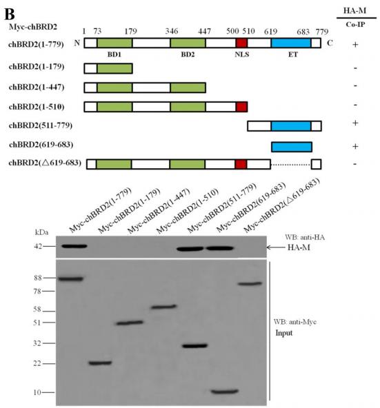
Figure 2| Characterization of the binding domains between the NDV M protein and chBRD2 protein. B Mapping the binding domain of the chBRD2 protein for the NDV M protein. Upper panel, schematic representation of the full-length and truncation mutants of the Myc-tagged chBRD2 protein. Lower panel, Myc-tagged chBRD2 or its truncation mutants (Input) interacting with HA-M was detected by western blot.
4). Schizosaccharomyces pombe MAP kinase Sty1 promotes survival of Δppr10 cells with defective mitochondrial protein synthesis. The international journal of biochemistry & cell biology, 2022
(PubMed: 36174923)
[IF=3.4]
Restrictive clause
Affinity Biosciences tests all products strictly. Citations are provided as a resource for additional applications that have not been validated by Affinity Biosciences. Please choose the appropriate format for each application and consult Materials and Methods sections for additional details about the use of any product in these publications.
For Research Use Only.
Not for use in diagnostic or therapeutic procedures. Not for resale. Not for distribution without written consent. Affinity Biosciences will not be held responsible for patent infringement or other violations that may occur with the use of our products. Affinity Biosciences, Affinity Biosciences Logo and all other trademarks are the property of Affinity Biosciences LTD.


