製品: beta-Tubulin Antibody
カタログ: T0023
タンパク質の説明: Mouse monoclonal antibody to beta-Tubulin
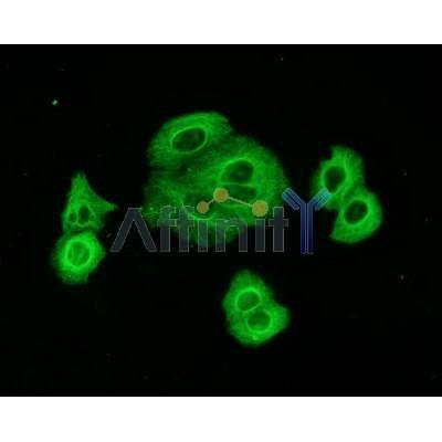
アプリケーション: WB IHC IF/ICC ELISA
反応性: Human, Mouse, Rat, Sheep, Rabbit, Dog, Monkey, Hamster, Chicken
分子量: 55KD; 50kD(Calculated).
ユニプロット: P07437
RRID: AB_2813772

類似製品を見る>>

   サイズ 価格 在庫状況
 50ul $150 在庫あり
 100ul $250 在庫あり
 200ul $350 在庫あり
 1ml $1200 在庫あり

リードタイム: 当日配達

For pricing and ordering contact:
お問い合わせ先

製品説明

ソース:
Mouse
アプリケーション:
WB 1:10000-1:500000, IHC 1:50-1:200, IF/ICC: 1:200
*The optimal dilutions should be determined by the end user.
*Tips:

WB: For western blot detection of denatured protein samples. IHC: For immunohistochemical detection of paraffin sections (IHC-p) or frozen sections (IHC-f) of tissue samples. IF/ICC: For immunofluorescence detection of cell samples. ELISA(peptide): For ELISA detection of antigenic peptide.

反応性:
Human,Mouse,Rat,Sheep,Rabbit,Dog,Monkey,Hamster,Chicken
クローナリティ:
Monoclonal [AFfirm039]
特異性:
beta-Tubulin Mouse Monoclonal antibody detects endogenous levels of total beta-Tubulin protein.
RRID:
AB_2813772
引用形式: Affinity Biosciences Cat# T0023, RRID:AB_2813772.
コンジュゲート:
Unconjugated.
精製:
Affinity-chromatography.
保存:
Mouse IgG1 in phosphate buffered saline (without Mg2+ and Ca2+), pH 7.4, 150mM NaCl. Store at -20 °C. Stable for 12 months from date of receipt.
別名:

折りたたみ/展開

TUBB3, CDCBM, Beta III Tubulin, Class III beta-tubulin, TUBB4, Tubulin, beta 3, Tubulin beta-III, Tubulin beta-3 chain, Tubulin beta-4 chain, Tubulin, beta 3 class III, CFEOM3A

免疫原

免疫原:

Full-length beta-tubulin protein of human.

Uniprot:
遺伝子(ID):
発現特異性:
P07437 TBB5_HUMAN:

Ubiquitously expressed with highest levels in spleen, thymus and immature brain.

タンパク質の説明:
Microtubules are constituent parts of the mitotic apparatus, cilia, flagella, and elements of the cytoskeleton. They consist principally of 2 soluble proteins, alpha- and beta-tubulin, each of about 55,000 kDa. Antibodies against beta Tubulin are useful as loading controls for Western Blotting. However it should be noted that levels of beta-Tubulin may not be stable in certain cells. For example, expression of beta-Tubulin in adipose tissue is very low and therefore beta-Tubulin should not be used as loading control for these tissues.
タンパク質配列:
MREIVHIQAGQCGNQIGAKFWEVISDEHGIDPTGTYHGDSDLQLDRISVYYNEATGGKYVPRAILVDLEPGTMDSVRSGPFGQIFRPDNFVFGQSGAGNNWAKGHYTEGAELVDSVLDVVRKEAESCDCLQGFQLTHSLGGGTGSGMGTLLISKIREEYPDRIMNTFSVVPSPKVSDTVVEPYNATLSVHQLVENTDETYCIDNEALYDICFRTLKLTTPTYGDLNHLVSATMSGVTTCLRFPGQLNADLRKLAVNMVPFPRLHFFMPGFAPLTSRGSQQYRALTVPELTQQVFDAKNMMAACDPRHGRYLTVAAVFRGRMSMKEVDEQMLNVQNKNSSYFVEWIPNNVKTAVCDIPPRGLKMAVTFIGNSTAIQELFKRISEQFTAMFRRKAFLHWYTGEGMDEMEFTEAESNMNDLVSEYQQYQDATAEEEEDFGEEAEEEA

PTMs - P07437 基板として

Site PTM Type Enzyme
R2 Methylation
C12 S-Nitrosylation
K19 Acetylation
K19 Methylation
K19 Ubiquitination
S25 Phosphorylation
T33 Phosphorylation
T35 Phosphorylation
Y36 Phosphorylation
S40 Phosphorylation
R46 Methylation
S48 Phosphorylation
Y50 Phosphorylation
Y51 Phosphorylation
T55 Phosphorylation
K58 Acetylation
K58 Methylation
K58 Sumoylation
K58 Ubiquitination
Y59 Phosphorylation
T72 Phosphorylation
S75 Phosphorylation
S78 Phosphorylation
S95 Phosphorylation
K103 Acetylation
K103 Sumoylation
K103 Ubiquitination
Y106 Phosphorylation
T107 Phosphorylation
S115 Phosphorylation
K122 Ubiquitination
S126 Phosphorylation
T136 Phosphorylation
S138 Phosphorylation
T143 Phosphorylation
S145 Phosphorylation
T149 Phosphorylation
S153 Phosphorylation
K154 Ubiquitination
Y159 Phosphorylation
R162 Methylation
T166 Phosphorylation
S168 Phosphorylation
S172 Phosphorylation
K174 Ubiquitination
Y183 Phosphorylation
T196 Phosphorylation
T199 Phosphorylation
Y200 Phosphorylation
Y208 Phosphorylation
K216 Ubiquitination
T218 Phosphorylation
T219 Phosphorylation
T221 Phosphorylation
Y222 Phosphorylation
S234 Phosphorylation
C239 S-Nitrosylation
K252 Sumoylation
K252 Ubiquitination
T274 Phosphorylation
S275 Phosphorylation Q5TCY1 (TTBK1)
R276 Methylation
S278 Phosphorylation
Y281 Phosphorylation
T285 Phosphorylation
T290 Phosphorylation
K297 Ubiquitination
C303 S-Nitrosylation
Y310 Phosphorylation
T312 Phosphorylation
R318 Methylation
S322 Phosphorylation
K324 Acetylation
K324 Sumoylation
K324 Ubiquitination
K336 Acetylation
K336 Ubiquitination
S338 Phosphorylation
S339 Phosphorylation
Y340 Phosphorylation
K350 Sumoylation
K350 Ubiquitination
T351 Phosphorylation
K362 Ubiquitination
T366 Phosphorylation
S371 Phosphorylation
K379 Acetylation
K379 Ubiquitination
S382 Phosphorylation
T386 Phosphorylation
K392 Ubiquitination
T399 Phosphorylation
Y422 Phosphorylation
Y425 Phosphorylation
T429 Phosphorylation

研究背景

機能:

Tubulin is the major constituent of microtubules. It binds two moles of GTP, one at an exchangeable site on the beta chain and one at a non-exchangeable site on the alpha chain.

PTMs:

Some glutamate residues at the C-terminus are polyglutamylated, resulting in polyglutamate chains on the gamma-carboxyl group. Polyglutamylation plays a key role in microtubule severing by spastin (SPAST). SPAST preferentially recognizes and acts on microtubules decorated with short polyglutamate tails: severing activity by SPAST increases as the number of glutamates per tubulin rises from one to eight, but decreases beyond this glutamylation threshold.

Some glutamate residues at the C-terminus are monoglycylated but not polyglycylated due to the absence of functional TTLL10 in human. Monoglycylation is mainly limited to tubulin incorporated into axonemes (cilia and flagella). Both polyglutamylation and monoglycylation can coexist on the same protein on adjacent residues, and lowering glycylation levels increases polyglutamylation, and reciprocally. The precise function of monoglycylation is still unclear (Probable).

Phosphorylated on Ser-172 by CDK1 during the cell cycle, from metaphase to telophase, but not in interphase. This phosphorylation inhibits tubulin incorporation into microtubules.

細胞の位置付け:

Cytoplasm>Cytoskeleton.

Extracellular region or secreted Cytosol Plasma membrane Cytoskeleton Lysosome Endosome Peroxisome ER Golgi apparatus Nucleus Mitochondrion Manual annotation Automatic computational assertionSubcellular location
組織特異性:

Ubiquitously expressed with highest levels in spleen, thymus and immature brain.

サブユニット構造:

Heterodimer of alpha and beta chains. A typical microtubule is a hollow water-filled tube with an outer diameter of 25 nm and an inner diameter of 15 nM. Alpha-beta heterodimers associate head-to-tail to form protofilaments running lengthwise along the microtubule wall with the beta-tubulin subunit facing the microtubule plus end conferring a structural polarity. Microtubules usually have 13 protofilaments but different protofilament numbers can be found in some organisms and specialized cells. Interacts with PIFO. Interacts with DIAPH1. Interacts with MX1 (By similarity). May interact with RNABP10 (By similarity). Interacts with CFAP157 (By similarity).

タンパク質ファミリー:

The highly acidic C-terminal region may bind cations such as calcium.

Belongs to the tubulin family.

研究領域

· Cellular Processes > Transport and catabolism > Phagosome.   (View pathway)

· Cellular Processes > Cellular community - eukaryotes > Gap junction.   (View pathway)

· Human Diseases > Infectious diseases: Bacterial > Pathogenic Escherichia coli infection.

参考文献

1). The clinical antiprotozoal drug nitazoxanide and its metabolite tizoxanide extend Caenorhabditis elegans lifespan and healthspan. Acta pharmaceutica Sinica. B, 2024 (PubMed: 39027239) [IF=14.5]

2). ALKBH5 regulates cardiomyocyte proliferation and heart regeneration by demethylating the mRNA of YTHDF1. Theranostics, 2021 (PubMed: 33456585) [IF=12.4]

Application: WB    Species: mouse    Sample: cardiomyocytes

Figure 8.| ALKBH5 upregulates the mRNA stability of YTHDF1 in turn promotes the translation of YAP. (A) m6A-meRIP-qPCR of YTHDF1 in CTL or ALKBH5 OE P1 CMs (***P < 0.001, n = 4). (B) RT-qPCR analysis of YTHDF1 in cultured P1 cardiomyocytes transfected with control or ALKBH5 OE plasmid (**P < 0.01, n = 4). (C) RT-qPCR analysis of YTHDF1 in cultured P1 cardiomyocytes transfected with control siRNA or ALKBH5 siRNAs (*P < 0.05, n = 4). (D) Western blot analysis of ALKBH5 and YTHDF1 in cultured P1 cardiomyocytes transfected with control or ALKBH5 OE plasmid.

3). Free CA125 promotes ovarian cancer cell migration and tumor metastasis by binding Mesothelin to reduce DKK1 expression and activate the SGK3/FOXO3 pathway. International Journal of Biological Sciences, 2023 (PubMed: 33613114) [IF=9.2]

Application: WB    Species: Human    Sample: A2780 and OVCAR3 cells

Figure 2 CA125 downregulates DKK1 expression in ovarian cancer cells. qPCR (A-B), ELISA (C-D), and Western blot (E-F) analysis of DKK1 mRNA levels in A2780 and OVCAR3 cells treated with CA125 at 0, 0.2. and 0.4 µg/mL for 48 h. The results represent the mean ± SD. *p<0.05, **p<0.01, ****p<0.0001.

4). EGFR tyrosine kinase activity and Rab GTPases coordinate EGFR trafficking to regulate macrophage activation in sepsis. Cell Death & Disease, 2022 (PubMed: 36344490) [IF=9.0]

5). Combination of resolvin E1 and lipoxin A4 promotes the resolution of pulpitis by inhibiting NF-κB activation through upregulating sirtuin 7 in dental pulp fibroblasts. CELL PROLIFERATION, 2022 (PubMed: 35411569) [IF=8.5]

Application: WB    Species: rat    Sample: Dental pulp fibroblasts

FIGURE 1 |Effects of combined administration of RvE1 and LXA4 on pro-inflammatory factor expression.(C) NLRP3, (D) caspase-1, (E) IL-1β and (F) IL-18 mRNA levels on LPS-induced DPFs detected by qPCR and (G) their protein levels tested by western blotting (normalized to that of β-tubulin).

6). Harmine is an effective therapeutic small molecule for the treatment of cardiac hypertrophy. Acta Pharmacologica Sinica, 2021 (PubMed: 33785860) [IF=8.2]

Application: WB    Species: Rat    Sample: cardiac tissues

Fig. 8: Harmine attenuates inflammatory response and NF-κB activation both in vivo and vitro. a qPCR analyses of inflammatory-related genes mRNA levels in rat cardiac tissues (normalized to 18S expression). b Representative Western blot analysis of phosphorylated NF-κB p65 and NF-κB p65 protein expression in the heart tissues of WKYs and SHRs after 3-month treatment. c–e Quantification of phosphorylated NF-κB p65 and NF-κB p65 protein expression normalized to β-Tubulin, and phosphorylated NF-κB p65/NF-κB p65 ratio by densitometric analysis. f Representative images of hESC-derived cardiomyocytes stained with cTnT (green) antibody. The hESC-derived cardiomyocytes were treated with LPS (100 ng/mL) for 3 days or were pre-treated with harmine (10 μM) for 3 h and then co-treated with LPS (100 ng/mL) for 3 days (n > 200 per group). Scale bars, 100 μm. g Quantification of cell size changes in each group. h-i Representative immunoblots of phosphorylated NF-κB p65 (p-NF-κB p65) and NF-κB p65 in hESC-derived cardiomyocytes following treatment of LPS (10 μg/mL) for 3 h, or TNF-α (25;ng/mL) for 1 h and 3 h (upper), and quantification of relative protein expression by densitometry (bottom). In all harmine-treated group, hESC-derived cardiomyocytes were pre-treated with harmine (25 μM) for 0.5 h before LPS or TNF-α addition. β-Actin was used as loading control. Data are expressed as mean ± SEM. *P 

7). Methyl ferulic acid ameliorates alcohol-induced hepatic insulin resistance via miR-378b-mediated activation of PI3K-AKT pathway. BIOMEDICINE & PHARMACOTHERAPY, 2022 (PubMed: 34844105) [IF=7.5]

8). MagT1 regulated the odontogenic differentiation of BMMSCs induced byTGC-CM via ERK signaling pathway. Stem Cell Research & Therapy, 2019 (PubMed: 30704530) [IF=7.5]

Application: WB    Species: rat    Sample: BMMSCs

Fig. 5 | MagT1 was involved in the odontogenic differentiation of BMMSCs.aImmunocytochemistry showed protein expression of MagT1 in BMMSCs. b The protein expression of MagT1 was increased in BMMSCs during odontogenic differentiation. c MagT1-shRNA lentivirus inhibited the mRNA and protein expression of MagT1 in BMMSCs. d The protein levels of ALP, DSP, and DMP-1 were decreased in MagT1-shRNA BMMSCs during odontogenic differentiation after 14d. e In accordance, the extent of mineralization nodules was also significantly decreased.Scale bar = 100 μm

9). Human umbilical cord mesenchymal stem cells-derived exosomes for treating traumatic pancreatitis in rats. Stem Cell Research & Therapy, 2022 (PubMed: 35619158) [IF=7.5]

10). Asiatic acid alleviates cisplatin-induced renal fibrosis in tumor-bearing mice by improving the TFEB-mediated autophagy-lysosome pathway. Biomedicine & Pharmacotherapy, 2023 (PubMed: 37413899) [IF=7.5]

Application: WB    Species: Human    Sample: HK-2 cells

Fig. 1. AA treatment suppressed renal fibrosis induced by long-term cisplatin injection in nude mice suffering from tumors. (A) Representative images of Sirius red staining and the expression of α-SMA, Collagen I, and Fibronectin in the kidneys of tumor-bearing mice. (B-F) Western blotting and quantitative analysis of the renal expression of Collagen I, Collagen IV, α-SMA, TGF-β1 in tumor-bearing mice. (G-M) Western blotting and quantitative analysis of the expression of Fibronectin, Collagen I, Collagen IV, α-SMA, and TGF-β1 in HK-2 cells. (N-L) RT-PCR detected the renal expression of CTGF and TGF-β1. Each bar represents the mean±SEM from at least three independent experiments. Scale bars= 50 µm. Versus control * P 

もっと読みます

Restrictive clause


Affinity Biosciences tests all products strictly. Citations are provided as a resource for additional applications that have not been validated by Affinity Biosciences. Please choose the appropriate format for each application and consult Materials and Methods sections for additional details about the use of any product in these publications.

For Research Use Only.
Not for use in diagnostic or therapeutic procedures. Not for resale. Not for distribution without written consent. Affinity Biosciences will not be held responsible for patent infringement or other violations that may occur with the use of our products. Affinity Biosciences, Affinity Biosciences Logo and all other trademarks are the property of Affinity Biosciences LTD.