製品: CD81 Antibody
カタログ: DF2306
タンパク質の説明: Rabbit polyclonal antibody to CD81
アプリケーション: WB IHC IF/ICC
Cited expt.: WB
反応性: Human, Mouse, Rat
予測: Pig, Bovine, Horse, Sheep, Dog
分子量: 26 kDa; 26kD(Calculated).
ユニプロット: P60033
RRID: AB_2839530

類似製品を見る>>

   サイズ 価格 在庫状況
 100ul $280 在庫あり
 200ul $350 在庫あり

リードタイム: 当日配達

For pricing and ordering contact:
お問い合わせ先

製品説明

ソース:
Rabbit
アプリケーション:
WB 1:500-1:2000, IF/ICC 1:100-1:500, IHC 1:50-1:200
*The optimal dilutions should be determined by the end user. For optimal experimental results, antibody reuse is not recommended.
*Tips:

WB: For western blot detection of denatured protein samples. IHC: For immunohistochemical detection of paraffin sections (IHC-p) or frozen sections (IHC-f) of tissue samples. IF/ICC: For immunofluorescence detection of cell samples. ELISA(peptide): For ELISA detection of antigenic peptide.

反応性:
Human,Mouse,Rat
予測:
Pig(92%), Bovine(92%), Horse(100%), Sheep(92%), Dog(92%)
クローナリティ:
Polyclonal
特異性:
CD81 Antibody detects endogenous levels of total CD81.
RRID:
AB_2839530
引用形式: Affinity Biosciences Cat# DF2306, RRID:AB_2839530.
コンジュゲート:
Unconjugated.
精製:
The antiserum was purified by peptide affinity chromatography using SulfoLink™ Coupling Resin (Thermo Fisher Scientific).
保存:
Rabbit IgG in phosphate buffered saline, pH 7.4, 150mM NaCl, 0.02% sodium azide and 50% glycerol. Store at -20 °C. Stable for 12 months from date of receipt.
別名:

折りたたみ/展開

26 kDa cell surface protein TAPA 1; 26 kDa cell surface protein TAPA-1; 26 kDa cell surface protein TAPA1; CD 81; CD81; CD81 antigen (target of antiproliferative antibody 1); CD81 antigen; CD81 molecule; CD81_HUMAN; CVID6; S5.7; TAPA 1; TAPA1; Target of the antiproliferative antibody 1; Tetraspanin 28; Tetraspanin-28; Tetraspanin28; Tspan 28; Tspan-28; Tspan28;

免疫原

免疫原:

A synthesized peptide derived from human CD81, corresponding to a region within the internal amino acids.

Uniprot:
遺伝子(ID):
発現特異性:
P60033 CD81_HUMAN:

Expressed on B cells (at protein level) (PubMed:20237408). Expressed in hepatocytes (at protein level) (PubMed:12483205). Expressed in monocytes/macrophages (at protein level) (PubMed:12796480). Expressed on both naive and memory CD4-positive T cells (at protein level) (PubMed:22307619).

タンパク質の説明:
May play an important role in the regulation of lymphoma cell growth. Interacts with a 16-kDa Leu-13 protein to form a complex possibly involved in signal transduction. May acts a the viral receptor for HCV
タンパク質配列:
MGVEGCTKCIKYLLFVFNFVFWLAGGVILGVALWLRHDPQTTNLLYLELGDKPAPNTFYVGIYILIAVGAVMMFVGFLGCYGAIQESQCLLGTFFTCLVILFACEVAAGIWGFVNKDQIAKDVKQFYDQALQQAVVDDDANNAKAVVKTFHETLDCCGSSTLTALTTSVLKNNLCPSGSNIISNLFKEDCHQKIDDLFSGKLYLIGIAAIVVAVIMIFEMILSMVLCCGIRNSSVY

種類予測

種類予測:

Score>80(red) has high confidence and is suggested to be used for WB detection. *The prediction model is mainly based on the alignment of immunogen sequences, the results are for reference only, not as the basis of quality assurance.

Species
Results
Score
Horse
100
Pig
92
Bovine
92
Sheep
92
Dog
92
Chicken
40
Xenopus
0
Zebrafish
0
Rabbit
0
Model Confidence:
High(score>80) Medium(80>score>50) Low(score<50) No confidence

研究背景

機能:

Structural component of specialized membrane microdomains known as tetraspanin-enriched microdomains (TERMs), which act as platforms for receptor clustering and signaling. Essential for trafficking and compartmentalization of CD19 receptor on the surface of activated B cells. Upon initial encounter with microbial pathogens, enables the assembly of CD19-CR2/CD21 and B cell receptor (BCR) complexes at signaling TERMs, lowering the threshold dose of antigen required to trigger B cell clonal expansion and antibody production. In T cells, facilitates the localization of CD247/CD3 zeta at antigen-induced synapses with B cells, providing for costimulation and polarization toward T helper type 2 phenotype. Present in MHC class II compartments, may also play a role in antigen presentation. Can act both as positive and negative regulator of homotypic or heterotypic cell-cell fusion processes. Positively regulates sperm-egg fusion and may be involved in acrosome reaction (By similarity). In myoblasts, associates with CD9 and PTGFRN and inhibits myotube fusion during muscle regeneration (By similarity). In macrophages, associates with CD9 and beta-1 and beta-2 integrins, and prevents macrophage fusion into multinucleated giant cells specialized in ingesting complement-opsonized large particles. Also prevents the fusion of mononuclear cell progenitors into osteoclasts in charge of bone resorption (By similarity). May regulate the compartmentalization of enzymatic activities. In T cells, defines the subcellular localization of dNTPase SAMHD1 and permits its degradation by the proteasome, thereby controlling intracellular dNTP levels. Also involved in cell adhesion and motility. Positively regulates integrin-mediated adhesion of macrophages, particularly relevant for the inflammatory response in the lung (By similarity).

(Microbial infection) Acts as a receptor for hepatitis C virus (HCV) in hepatocytes. Association with CLDN1 and the CLDN1-CD81 receptor complex is essential for HCV entry into host cell.

(Microbial infection) Involved in SAMHD1-dependent restriction of HIV-1 replication. May support early replication of both R5- and X4-tropic HIV-1 viruses in T cells, likely via proteasome-dependent degradation of SAMHD1.

(Microbial infection) Specifically required for Plasmodium falciparum infectivity of hepatocytes, controlling sporozoite entry into hepatocytes via the parasitophorous vacuole and subsequent parasite differentiation to exoerythrocytic forms.

PTMs:

Not glycosylated.

Likely constitutively palmitoylated at low levels. Protein palmitoylation is up-regulated upon coligation of BCR and CD9-C2R-CD81 complexes in lipid rafts.

細胞の位置付け:

Cell membrane>Multi-pass membrane protein. Basolateral cell membrane>Multi-pass membrane protein.
Note: Associates with CLDN1 and the CLDN1-CD81 complex localizes to the basolateral cell membrane.

Extracellular region or secreted Cytosol Plasma membrane Cytoskeleton Lysosome Endosome Peroxisome ER Golgi apparatus Nucleus Mitochondrion Manual annotation Automatic computational assertionSubcellular location
組織特異性:

Expressed on B cells (at protein level). Expressed in hepatocytes (at protein level). Expressed in monocytes/macrophages (at protein level). Expressed on both naive and memory CD4-positive T cells (at protein level).

タンパク質ファミリー:

Binds cholesterol in a cavity lined by the transmembrane spans.

Belongs to the tetraspanin (TM4SF) family.

研究領域

· Human Diseases > Infectious diseases: Parasitic > Malaria.

· Human Diseases > Infectious diseases: Viral > Hepatitis C.

· Organismal Systems > Immune system > B cell receptor signaling pathway.   (View pathway)

参考文献
1). Neuronal Extracellular Vesicles Carrying APOE Downregulate Filament Actin Polymerization Signaling to Inhibit Synapse Formation in Alzheimer's Disease. Journal of extracellular vesicles, 2026 (PubMed: 41806350) [IF=16.0]

2). Neddylation of Coro1a determines the fate of multivesicular bodies and biogenesis of extracellular vesicles. Journal of Extracellular Vesicles, 2021 (PubMed: 34623756) [IF=16.0]

Application: WB    Species: Mice    Sample: HEK293 cells

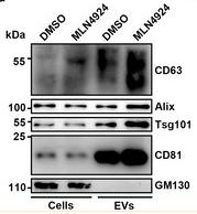
FIGURE 1 Neddylation inhibits EV secretion. (a) Flow cytometric analysis of EVs in the supernatants of HEK293 cells treated with DMSO or 100 nM MLN4924 for 12 h. Left, representative dot plots showing CD9 and CD81 staining of EVs captured with anti‑CD63‑coated beads after incubation with cell culture supernatants. Right, the ratio of CD9+ and CD81+ EVs. (b–d) EVs were purified from equal numbers of HEK293 cells treated with DMSO or 100 nM MLN4924 for 12 h. The BCA assay was used to determine the amount of EV protein (b). NTA to determine the EV concentration (c). WB analysis to detect the indicated EV markers (d). (e) WB analysis to detect the indicated proteins in HEK293 cells overexpressing NEDD8. (f) Flow cytometric ratio of CD9+ EVs in the supernatants of HEK293 cells overexpressing NEDD8. (g) ELISA analysis of CD9+ and CD81+ EVs in sera of mice intraperitoneally injected with MLN4924 at the indicated dose for 72 h. Representative results from three independent experiments are shown. n, sample number; *P < 0.05; **P < 0.01; ***P < 0.001 (unpaired two‐tailed Student's t‐test except for one‐way ANOVA followed by Tukey test in G; mean ± s.d.)

3). Proteomic insights uncover enhanced neurotherapeutic potential in conditioned mesenchymal stem cell-derived extracellular vesicles. Extracellular Vesicle, 2024 [IF=16.0]

Application: WB    Species: Mouse    Sample:

Fig. 1. Characterization of extracellular vesicles (EVs).(A–C) Western blots of membrane protein CD9, CD81, TSG 101 and cytoplasmic protein Calnexin in MSC-EVs (A), Conditioned (Con)1 EVs (B) and Con2 EVs (C), while these proteins expressed in MSCs were set as the control (A–C); (B) transmission electron microscopy (TEM) displays the typical morphology of MSC-EVs (D) and conditioned EVs (E and F). Bar = 100 nm; (G–I) nanoparticle tracking analysis (NTA) presents the size distribution of each type of EVs.

4). Iron Oxide Nanoparticles Engineered Macrophage-Derived Exosomes for Targeted Pathological Angiogenesis Therapy. ACS nano, 2024 (PubMed: 38412252) [IF=15.8]

Application: WB    Species: Mouse    Sample:

Figure 2 Characterization of ESIONPs@EXO derived from ESIONPs engineered macrophages. (A) The morphology of EXO and ESIONPs@EXO determined by TEM. Scale bar: 200 nm (left) and 100 nm (right). (B) The size distribution of EXO and ESIONPs@EXO evaluated by NTA. (C) Western blot analysis of CD9, CD63, CD81, TSG101, and calnexin. (D) Relaxation properties of ESIONPs@EXO. (E) T1 and T2 weighted MR images of ESIONPs@EXO at different concentrations (measured on a 3 T MR scanner). 1/T1 (F) and 1/T2 (G) relaxation rates of ESIONPs@EXO at different concentrations.

5). Intranasal delivery of engineered extracellular vesicles loaded with miR-206-3p antagomir ameliorates Alzheimer's disease phenotypes. Theranostics, 2024 (PubMed: 39659569) [IF=12.4]

Application: WB    Species: Mouse    Sample:

Figure 1. Preparation and characterization of MSC-derived extracellular vesicles loaded with miR-206-3p antagomir. (A) Schema illustrates the process of constructing engineered MSC-EVs through electroporation. (B) Total RNA was extracted from engineered MSC-EVs and quantified. The RNA content in MSC-EVs was evaluated under different treatment conditions, n = 4. (C-E) Representative size distribution (C-D) and TEM images (E) of MSC-EVs and MSC-EVs-anta. (F) The expression of CD9, CD63, CD81, TSG101, β-actin and negative marker Calnexin in naive MSC-EVs, electroporated MSC-EVs, and MSC-EVs-anta was determined by immunoblotting.

6). Extracellular vesicle-packaged GBP2 from macrophages aggravates sepsis-induced acute lung injury by promoting ferroptosis in pulmonary vascular endothelial cells. Redox biology, 2025 (PubMed: 40156957) [IF=10.7]

Application: WB    Species: Mouse    Sample:

Fig. 2. SMφ-derived EVs induce ferroptosis in pulmonary microvascular endothelial cells. (A) The flowchart of EV enrichment by ultracentrifugation. (B) Transmission electron microscopy (TEM) images of EVs isolated from the medium of Mφ and SMφ. Scale bar = 100 nm. (C) Representative nanoparticle tracking analysis images showing the distribution and average diameter of EVs. (D) Representative immunoblotting of EV markers. (E) Representative immunofluorescence staining of Hoechst/CFSE after co-culture with human pulmonary microvascular endothelial cells (HPMECs) using CFSE-labeled EVs. Scale bar = 10 μm. (F) Quantification of CCK8 assays performed on HPMECs co-cultured with Mφ-EVs and SMφ-EVs (n = 5). (G) Representative images and quantification of the Zombie NIR™ staining in HPMECs co-cultured with Mφ-EVs and SMφ-EVs in the presence or absence of ferrostatin-1 (Fer-1) (n = 3). (H) Representative immunofluorescence staining for DAPI/CD31/ZO-1 and DAPI/CD31/occludin in HPMECs. Scale bar = 50 μm. (I) Representative immunoblotting and quantification of barrier-associated proteins (ZO-1, VE-Cadherin and occludin) in HPMECs (n = 3). (J) Representative images of mitochondrial membrane potential of HPMECs. Scale bar = 50 μm. (K) Representative images of liperfluo staining in HPMECs. Scale bar = 50 μm. (L) C11-BODIPY assay evaluating the lipid ROS levels in HPMECs (n = 3). (M) Representative immunoblotting and quantification of SLC7A11 and GPX4 in HPMECs (n = 3). (N) Representative images of immunofluorescence staining for DAPI/CD31/SLC7A11 and DAPI/CD31/GPX4 in HPMECs. Scale bar = 50 μm. Data are presented as mean ± SD.

7). THBS1 in macrophage-derived exosomes exacerbates cerebral ischemia-reperfusion injury by inducing ferroptosis in endothelial cells. Journal of neuroinflammation, 2025 (PubMed: 39994679) [IF=9.3]

8). Injectable hydrogel microspheres delivering cartilage-targeted LGR5-engineered exosomes for osteoarthritis therapy. Materials today. Bio, 2025 (PubMed: 41560788) [IF=8.7]

9). Hypoxia Induces HIF-1α Activation in Tendon Stem Cells to Enhance Extracellular Vesicle-Mediated Tendon Repair. ACS applied materials & interfaces, 2025 (PubMed: 40675622) [IF=8.3]

10). Exosome from indoleamine 2,3-dioxygenase-overexpressing bone marrow mesenchymal stem cells accelerates repair process of ischemia/reperfusion-induced acute kidney injury by regulating macrophages polarization. Stem cell research & therapy, 2022 (PubMed: 35902956) [IF=7.5]

もっと読みます

Restrictive clause


Affinity Biosciences tests all products strictly. Citations are provided as a resource for additional applications that have not been validated by Affinity Biosciences. Please choose the appropriate format for each application and consult Materials and Methods sections for additional details about the use of any product in these publications.

For Research Use Only.
Not for use in diagnostic or therapeutic procedures. Not for resale. Not for distribution without written consent. Affinity Biosciences will not be held responsible for patent infringement or other violations that may occur with the use of our products. Affinity Biosciences, Affinity Biosciences Logo and all other trademarks are the property of Affinity Biosciences LTD.