製品: Cyclin E1 Antibody
カタログ: AF0144
タンパク質の説明: Rabbit polyclonal antibody to Cyclin E1
アプリケーション: WB IHC IF/ICC
反応性: Human, Mouse, Rat
分子量: 49kDa; 47kD(Calculated).
ユニプロット: P24864
RRID: AB_2833326

類似製品を見る>>

   サイズ 価格 在庫状況
 100ul $280 在庫あり
 200ul $350 在庫あり

リードタイム: 当日配達

For pricing and ordering contact:
お問い合わせ先

製品説明

ソース:
Rabbit
アプリケーション:
WB 1:500-1:3000, IHC 1:50-1:200, IF/ICC: 1:100-1:500
*The optimal dilutions should be determined by the end user.
*Tips:

WB: For western blot detection of denatured protein samples. IHC: For immunohistochemical detection of paraffin sections (IHC-p) or frozen sections (IHC-f) of tissue samples. IF/ICC: For immunofluorescence detection of cell samples. ELISA(peptide): For ELISA detection of antigenic peptide.

反応性:
Human,Mouse,Rat
クローナリティ:
Polyclonal
特異性:
Cyclin E1 Antibody detects endogenous levels of total Cyclin E1.
RRID:
AB_2833326
引用形式: Affinity Biosciences Cat# AF0144, RRID:AB_2833326.
コンジュゲート:
Unconjugated.
精製:
The antiserum was purified by peptide affinity chromatography using SulfoLink™ Coupling Resin (Thermo Fisher Scientific).
保存:
Rabbit IgG in phosphate buffered saline , pH 7.4, 150mM NaCl, 0.02% sodium azide and 50% glycerol. Store at -20 °C. Stable for 12 months from date of receipt.
別名:

折りたたみ/展開

CCNE; Ccne1; CCNE1_HUMAN; cyclin E variant ex5del; cyclin E variant ex7del; Cyclin E1; Cyclin Es; Cyclin Et; CyclinE; G1/S specific cyclin E; G1/S-specific cyclin-E1;

免疫原

免疫原:
Uniprot:
遺伝子(ID):
発現特異性:
P24864 CCNE1_HUMAN:

Highly expressed in testis and placenta. Low levels in bronchial epithelial cells.

タンパク質の説明:
CCNE1 a member of the highly conserved cyclin family, whose members are characterized by a dramatic periodicity in protein abundance through the cell cycle. Cyclins function as regulators of CDK kinases. Forms a complex with and functions as a regulatory subunit of CDK2, whose activity is required for cell cycle G1/S transition. Accumulates at the G1-S phase boundary and is degraded as cells progress through S phase. Two alternatively spliced isoforms have been described.
タンパク質配列:
MPRERRERDAKERDTMKEDGGAEFSARSRKRKANVTVFLQDPDEEMAKIDRTARDQCGSQPWDNNAVCADPCSLIPTPDKEDDDRVYPNSTCKPRIIAPSRGSPLPVLSWANREEVWKIMLNKEKTYLRDQHFLEQHPLLQPKMRAILLDWLMEVCEVYKLHRETFYLAQDFFDRYMATQENVVKTLLQLIGISSLFIAAKLEEIYPPKLHQFAYVTDGACSGDEILTMELMIMKALKWRLSPLTIVSWLNVYMQVAYLNDLHEVLLPQYPQQIFIQIAELLDLCVLDVDCLEFPYGILAASALYHFSSSELMQKVSGYQWCDIENCVKWMVPFAMVIRETGSSKLKHFRGVADEDAHNIQTHRDSLDLLDKARAKKAMLSEQNRASPLPSGLLTPPQSGKKQSSGPEMA

PTMs - P24864 基板として

Site PTM Type Enzyme
S59 Phosphorylation
S73 Phosphorylation P49841 (GSK3B)
T77 Phosphorylation P49841 (GSK3B)
S90 Phosphorylation
S103 Phosphorylation
S381 Phosphorylation
S387 Phosphorylation P24941 (CDK2)
S391 Phosphorylation
T395 Phosphorylation P49841 (GSK3B) , P24941 (CDK2) , P49840 (GSK3A)
S399 Phosphorylation P24941 (CDK2)

PTMs - P24864 酵素として

Substrate Site Source
Q8TDN4-2 (CABLES1) S48 Uniprot

研究背景

機能:

Essential for the control of the cell cycle at the G1/S (start) transition.

PTMs:

Phosphorylation of both Thr-395 by GSK3 and Ser-399 by CDK2 creates a high affinity degron recognized by FBXW7, and accelerates degradation via the ubiquitin proteasome pathway. Phosphorylation at Thr-77 creates a low affinity degron also recognized by FBXW7.

Ubiquitinated by UHRF2; appears to occur independently of phosphorylation.

細胞の位置付け:

Nucleus.

Extracellular region or secreted Cytosol Plasma membrane Cytoskeleton Lysosome Endosome Peroxisome ER Golgi apparatus Nucleus Mitochondrion Manual annotation Automatic computational assertionSubcellular location
組織特異性:

Highly expressed in testis and placenta. Low levels in bronchial epithelial cells.

サブユニット構造:

Interacts with CDK2 protein kinase to form a serine/threonine kinase holoenzyme complex. The cyclin subunit imparts substrate specificity to the complex. Found in a complex with CDK2, CABLES1 and CCNA1 (By similarity). Part of a complex consisting of UHRF2, CDK2 and CCNE1. Interacts directly with UHRF2; the interaction ubiquitinates CCNE1 and appears to occur independently of CCNE1 phosphorylation. Interacts with INCA1.

タンパク質ファミリー:

Belongs to the cyclin family. Cyclin E subfamily.

研究領域

· Cellular Processes > Cell growth and death > Cell cycle.   (View pathway)

· Cellular Processes > Cell growth and death > Oocyte meiosis.   (View pathway)

· Cellular Processes > Cell growth and death > p53 signaling pathway.   (View pathway)

· Cellular Processes > Cell growth and death > Cellular senescence.   (View pathway)

· Environmental Information Processing > Signal transduction > PI3K-Akt signaling pathway.   (View pathway)

· Human Diseases > Infectious diseases: Viral > Hepatitis B.

· Human Diseases > Infectious diseases: Viral > Measles.

· Human Diseases > Infectious diseases: Viral > Human papillomavirus infection.

· Human Diseases > Cancers: Overview > Pathways in cancer.   (View pathway)

· Human Diseases > Cancers: Overview > Viral carcinogenesis.

· Human Diseases > Cancers: Overview > MicroRNAs in cancer.

· Human Diseases > Cancers: Specific types > Prostate cancer.   (View pathway)

· Human Diseases > Cancers: Specific types > Small cell lung cancer.   (View pathway)

· Human Diseases > Cancers: Specific types > Gastric cancer.   (View pathway)

参考文献

1). TCA-phospholipid-glycolysis targeted triple therapy effectively suppresses ATP production and tumor growth in glioblastoma. Theranostics, 2023 (PubMed: 36276638) [IF=12.4]

Application: WB    Species: Human    Sample: TBD0220 and U87MG cells

Figure 3 Reduction in ATP production hinders cell proliferation and contributes to G1/S arrest in GBM. (A-B) The relative viability of TBD0220 (A) and U87MG (B) cells was measured using a CCK-8 kit (n = 3). (C-D) Colony formation assay to detect GBM cell growth (D), and the quantification of colony numbers (C) (n = 3). (E) Cell cycle analysis using flow cytometry after incubation with different treatments like EPIC (20 µM), AA (25 µM), or EPIC (20 µM) + AA (25 µM) for 48 h, and the results are plotted as a histogram (n = 3) (F) Representative western blotting showing the expression of p21 and Rb and their downstream targets. The results were normalized to Tubulin with the control group as 1. Protein expression was quantified by ImageJ. (G) Representative confocal images of CDK6 after the treatment with AA (25 µM), EPIC (20 µM), or EPIC (20 µM) + AA (25 µM) for 48 h (n = 6). (H) The quantitative analysis of fluorescence images of CDK6 (n = 6). All data are shown as the mean values ± SD, and p values are based on one-way or two-way ANOVA. ****p < 0.0001, ***p < 0.001, **p < 0.01, *p < 0.05. Scale bar = 20 μm.

2). Combination LSD1 and HOTAIR-EZH2 inhibition disrupts cell cycle processes and induces apoptosis in glioblastoma cells. PHARMACOLOGICAL RESEARCH, 2021 (PubMed: 34246782) [IF=9.3]

3). Epigallocatechin gallate suppresses mitotic clonal expansion and adipogenic differentiation of preadipocytes through impeding JAK2/STAT3-mediated transcriptional cascades. Phytomedicine : international journal of phytotherapy and phytopharmacology, 2024 (PubMed: 38552377) [IF=7.9]

4). Ganoderma lucidum polysaccharide inhibits HSC activation and liver fibrosis via targeting inflammation, apoptosis, cell cycle, and ECM-receptor interaction mediated by TGF-β/Smad signaling. Phytomedicine, 2023 (PubMed: 36603342) [IF=7.9]

5). Long non-coding RNA RP11-197K6.1 as ceRNA promotes colorectal cancer progression via miR-135a-5p/DLX5 axis. Journal of translational medicine, 2024 (PubMed: 38760791) [IF=7.4]

Application: WB    Species: Human    Sample: HCT116 and SW480 cells

Fig. 2 Functional effects of lncRNA RP11-197K6.1 knockdown in CRC cells. A: Confirmation of lncRNA RP11-197K6.1 knockdown efficiency in HCT116 cells by RT-qPCR. B: Reduction in the proliferation of HCT116 and SW480 cells post-knockdown, as determined by proliferation assays. C: Increase in the apoptosis of HCT116 and SW480 cells following lncRNA RP11-197K6.1 knockdown, as measured by flow cytometry. D & E: Decrease in the migration and invasion of HCT116 and SW480 cells post-knockdown, as assessed by wound healing (scale = 200 μm) and Transwell assays (scale = 50 μm), respectively. 2 F: Changes in the levels of proteins related to the proliferation, apoptosis, and migration of HCT116 and SW480 cells after lncRNA RP11-197K6.1 knockdown, as analyzed by western blotting assay (**P 

6). Chelerythrine chloride inhibits the progression of colorectal cancer by targeting cancer-associated fibroblasts through intervention with WNT10B/β-catenin and TGFβ2/Smad2/3 axis. Phytotherapy research : PTR, 2023 (PubMed: 37402476) [IF=7.2]

7). Silica nanoparticles cause spermatogenesis dysfunction in mice via inducing cell cycle arrest and apoptosis. ECOTOXICOLOGY AND ENVIRONMENTAL SAFETY, 2022 (PubMed: 35051769) [IF=6.8]

Application: WB    Species: mouse    Sample: testis

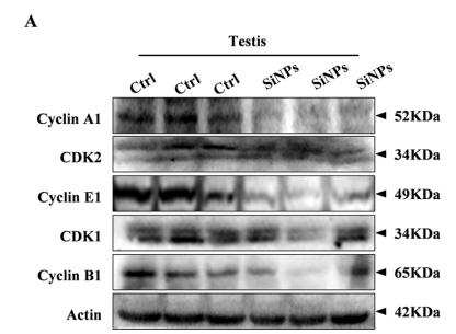
Fig. 5. |SiNPs affected the protein expressions of cell cycle regulators. (A) The protein expressions of cyclin A1, CDK2, cyclin E1, CDK1, and cyclin B1 were significantly decreased in the testis after exposure to SiNPs.

8). Loss of exosomal miR-200b-3p from hypoxia cancer-associated fibroblasts promotes tumorigenesis and reduces sensitivity to 5-Flourouracil in colorectal cancer via upregulation of ZEB1 and E2F3. Cancer Gene Therapy, 2023 (PubMed: 36890211) [IF=6.4]

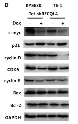
9). RECQL4 regulates DNA damage response and redox homeostasis in esophageal cancer. Cancer Biology & Medicine, 2021 (PubMed: 33628589) [IF=5.5]

Application: WB    Species: human    Sample: ESCC cells

Figure 4 |The loss of RECQL4 induces cell cycle arrest and cellular senescence. . (D) The protein levels of c-myc, p21, cyclin D, CDK6, cyclin E, Bax, and Bcl-2 were determined by Western blot in stable Tet-on inducible RECQL4 knockdown cell lines (KYSE30 and TE-1 cells) (+Dox) and controls (–Dox). Experiments were independently repeated 3 times. All data indicate the mean ±SD. *P < 0.05; **P < 0.01; ***P < 0.001.

Application: WB    Species: Human    Sample: KYSE30 and TE-1 cells

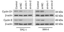
Figure 4 The loss of RECQL4 induces cell cycle arrest and cellular senescence. (A) Depletion of RECQL4 by siRNA. RECQL4 protein levels were measured by Western blot. KYSE30 and TE-1 cells were transfected with siRNA duplexes (200 nM) specific to RECQL4 or negative oligo in serum-free medium for 4 h, then replaced with complete medium for 24 h. Whole cell extracts were collected for Western blot analysis using RECQL4 antibodies. (B) Cell cycle distributions in RECQL4 knockdown cell lines (KYSE30 and TE-1 cells) and controls were determined by flow cytometry. (C) Cellular senescence was examined by SA-β-gal staining. Microscopic magnification (×200), Scale bar: 50 μm. (D) The protein levels of c-myc, p21, cyclin D, CDK6, cyclin E, Bax, and Bcl-2 were determined by Western blot in stable Tet-on inducible RECQL4 knockdown cell lines (KYSE30 and TE-1 cells) (+Dox) and controls (–Dox). Experiments were independently repeated 3 times. All data indicate the mean ± SD. *P < 0.05; **P < 0.01; ***P < 0.001.

10). OTUB1 promotes progression and proliferation of prostate cancer via deubiquitinating and stabling cyclin E1. Frontiers in Cell and Developmental Biology, 2021 (PubMed: 33537306) [IF=5.5]

Application: WB    Species: Mice    Sample: PC3 and C4-2 cells

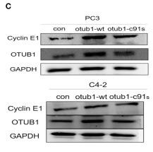
Figure 4 OTUB1 promotes and regulates the expression of Cyclin E1. (A) The relationship between OTUB1 and Cyclin E1 was analyzed via gene MANIA database. (B) The protein expression of Cyclin E1 in BPH, ADPC, and CRPC was detected by IHC staining. (C) Western blotting detecting the expression of Cyclin E1 in PC3 and C4-2 cells transfected with otub1 overexpression and otub1 c91s. (D) Western blotting detecting the expression of Cyclin E1 in PC3 and C4-2 cells transfected with OTUB1 siRNA. (E) Western blotting detecting the expression of Cyclin E1 in PC3 and C4-2 cells transfected with an increasing gradient OTUB1.

Application: IHC    Species: Mice    Sample: tumors

Figure 7 OTUB1 promotes the growth of PC3 cell via increasing Cyclin E1 in vivo. (A,B) PC3 cells transfected with OTUB1 overexpression and otub1 c91s were transplanted subcutaneously in nude mice. The effect of otub1 overexpression, otub1 c91s, and RO-3306 on the growth of PCa. (C) The tumor volume was measured with a caliper. (D) The tumor volumes were measured daily when the diameter grew into 2 mm. (E) IHC staining detecting the expression of OTUB1, ki-67, and Cyclin E1 in these tumors from nude mice. ****P < 0.0001.

もっと読みます

Restrictive clause


Affinity Biosciences tests all products strictly. Citations are provided as a resource for additional applications that have not been validated by Affinity Biosciences. Please choose the appropriate format for each application and consult Materials and Methods sections for additional details about the use of any product in these publications.

For Research Use Only.
Not for use in diagnostic or therapeutic procedures. Not for resale. Not for distribution without written consent. Affinity Biosciences will not be held responsible for patent infringement or other violations that may occur with the use of our products. Affinity Biosciences, Affinity Biosciences Logo and all other trademarks are the property of Affinity Biosciences LTD.